GeoMx Digital Spatial Profiler
A Flexible & Scalable Platform for Spatial Biology
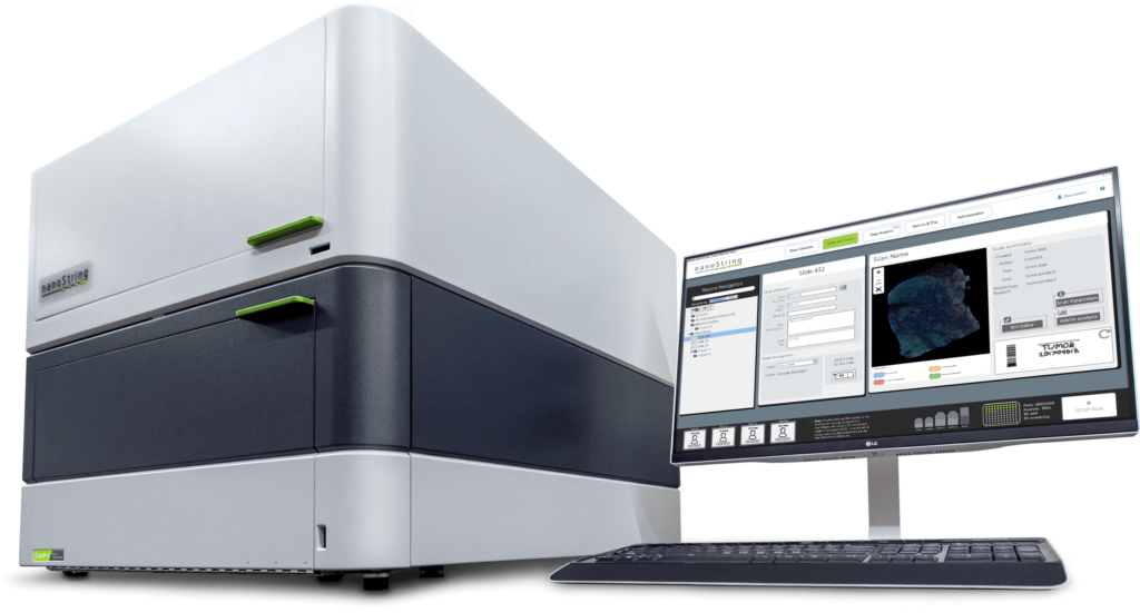
Quickly resolve tissue heterogeneity and the complexity of microenvironments with the GeoMx Digital Spatial Profiler (DSP), the most flexible and robust spatial multiomic platform for analysis of FFPE and fresh frozen tissue sections. GeoMx is the only spatial biology platform that non-destructively profiles expression of RNA and protein from distinct tissue compartments and cell populations with an automated and scalable workflow that integrates with standard histology staining.
What Can you do with GeoMx DSP?

Discover biomarkers predictive of therapeutic response
"Using two quantitative and spatially informed technologies in independent NSCLC cohorts, we show that CD44 overexpression in the tumor compartment, but not in the immune compartment, predicts clinical outcomes from PD-1 axis blockade." –Moutafi et al.
Profile the tumor microenvironment
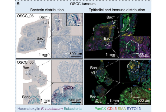
Spatial characterization of protein expression in the TME compartments infected with Fusobacterium species provided direct evidence of how bacteria may alter cells and hamper the immune response against cancer.
Uncover drug mechanism of action
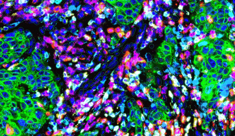
Explore the mechanism of actions and dose effects of an oncolytic viruses, CAR-T cells and combination therapy using Xenografts in laboratory animals
Spatial whole transcriptome profiling
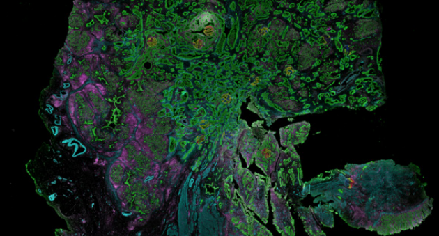
Spatial whole transcriptome profiling of pancreatic cancer redefines the molecular and cellular taxonomy associated with neoadjuvant therapy
GeoMx® Digital Spatial Profiler
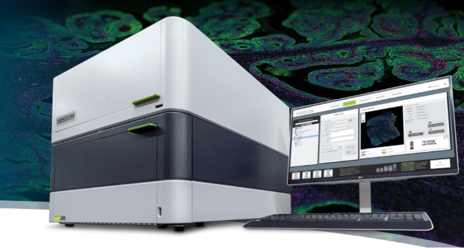
Spatially profile the whole transcriptome and 1200+ protein targets separately or simultaneously from your choice of sample inputs: whole tissue sections, tissue microarrays (TMAs), or organoids.
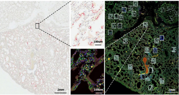
Unstand disease mechanisms and progression
Explore the heterogeneity of immune response to SARS-CoV2 help researchers identify two distinct phases of COVID-19
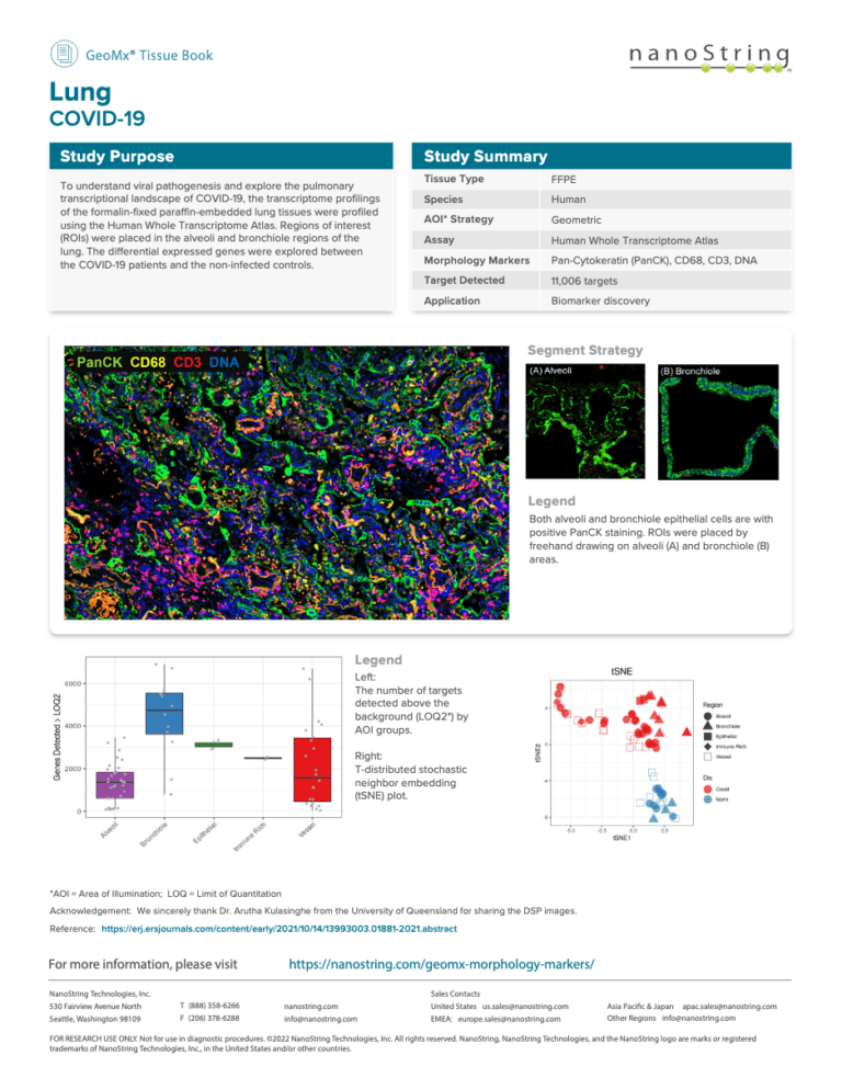
What is the Spatial Tissue Book?
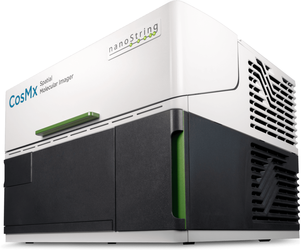
The Spatial Tissue Book is a repository of images, data, selection and segmentation strategies for different tissue types and diseases that have been analyzed through either the GeoMx® DSP Technology Access Program or the CosMx SMI Technology Access Program.
Each individual PDF page of the Tissue Book includes images of the tissue and is annotated by the disease(s) studied, species of interest, and type of assay performed.
Why Use the Tissue Book?
- See representative images for tissues and/or disease areas that you are studying.
- Get ideas on how to select ROIs and segment the tissue to meet your study design needs.
- Review spatial transcriptomics data from different tissues representing various diseases.
Explore the anatomy of a GeoMx tissue book page
- The type of tissue and disease profiled.
- Learn about the application and study design.
- Get a high-level overview of what kind of sample type was used, species, strategy for area of illumination as well as the assay and morphology markers used. Learn how many targets were detected from the assay for this particular study.
- See an actual image taken with either the GeoMx Digital Spatial Profiler or the CosMx Spatial Molecular Imager annotated with the different fluorescent markers used to outline tissue morphology and/or cell types.
- Learn more about the Segmentation Strategy and how individual regions of interest were compartmentalized into different areas of illumination and/or cell type populations.
- See how many targets were detected above the background for each tissue compartment and/or cell type profiled.
- View additional plots showing the results of the study.
- Here you can click on this link to visit our Morphology Marker page to learn about which fluorescently-tagged antibodies or RNAScope Probes have been used on the GeoMx DSP.
Tissue Book Collection
Browse entries here, or just download the complete tissue book.
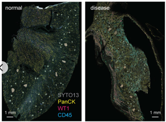
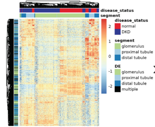
Find the Right Platform for Your Research
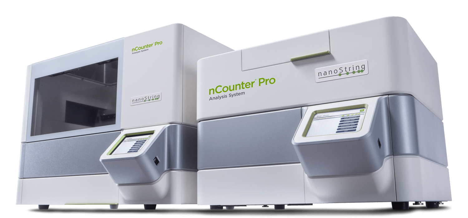
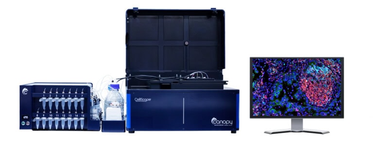
| Platform | CosMx | CellScape | GeoMx | nCounter | PaintScape |
|---|---|---|---|---|---|
| Applications | High fidelity, high sensitivity, subcellular imaging of the whole transcriptome with proteomics for complete pathway-level spatial insights | High fidelity, flexible subcellular proteomics for rapid and quantitative spatial phenotyping | High fidelity, unmatched discovery multiomics to profile from cells to tissue structures for spatial breakthroughs at scale | Rapid and robust targeted RNA and multiomic expression proven for translational and clinical studies | High fidelity, high precision multiplexed direct visualization of spatial organization of the 3D genome |
| Analyte | RNA, protein | Protein, RNA | RNA, protein | miRNA, Protein, RNA | DNA |
| Plex level | Target RNA assays (up to 6000 targets) Whole transcriptome RNA assays Up to 64 proteins | Targeted protein assays (up to 34 targets) Customizable assays up to 20 | Targeted RNA assays (up to 2000 targets) Whole transcriptome RNA assays Up to 1200 proteins | 800+ RNA 500+ protein | Targeted DNA Assays (419+ targets) |
| Resolution | Single-cell Subcellular | Single-cell Subcellular | Multicellular (RNA) Single-cell (protein) | Bulk | Single-nuclei |
| Sample compatibility | FFPE, FF, organoids, cultured cells | FFPE, FF, cell suspension, lysate | FFPE, FF | FFPE, FF, lysate | Human cell lines, FF tissue (coming soon) |
| Data readout | Imaging | Imaging | NGS, nCounter (Direct counting) | nCounter (Direct counting) | Imaging |